Nikon A1
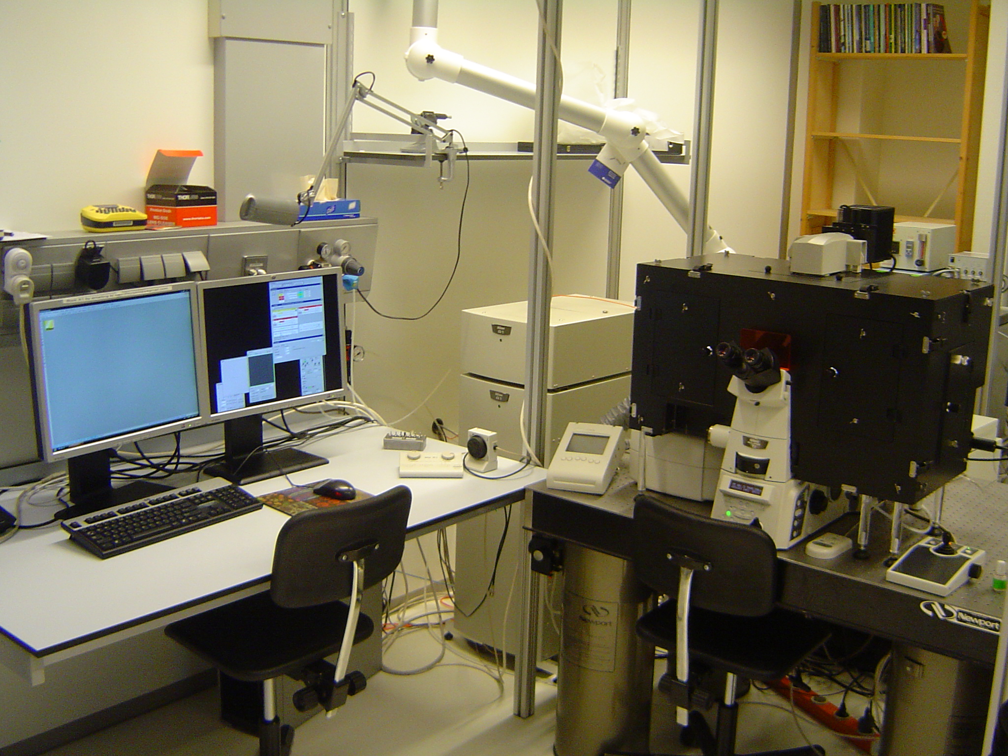 |
Location: | LCAM, room A2.34 |
| Contact person: |  |
|
| 2nd Contact person: |  |
|
| Techniques: | (Semi-)Widefield imaging Confocal imaging Standard confocal techniques like co-localization & FRAP Spatially Controled Illumination Microscopy (SCIM, previously CLEM) Spectral analysis (f.e. to study FRET) |
|
| Booking: | The A1 microscope can be booked by trained users only. To book the microscope, Ronald Breedijk (+7860) should be contacted. The composed measurement schedule will be published in the webcalender on Thursday, the week before. For first time-users contact Ronald Breedijk (+7860). | |
| Lasers: | 405 nm diode laser 440 nm diode laser 458 nm, Ar mW (CLEM unit) 488 nm, Ar mW 514 nm, Ar mW 561 nm, dpss mW 594 nm HeNe laser 640 nm, diode laser mW For more info about laser safety, contact Ronald Breedijk (+7860) |
|
| Description: The Nikon A1 confocal microscope for live-cell multi-clor imaging and with options to do fast imaging using the resonant scanner, SCIM microscopy or spectral analysis | Objectives: | 20x Plan Fluor, NA 0.75 (multi immersion) 40x Plan Fluor, NA 1.3 (oil) 60x Plan Apo VC, NA 1.4 (oil) |
| Microscope: | Nikon Ti + mercury and transmission lamp DAPI, GFP, FITC and TexasRed fluorescence cubes motorized xyz stage |
|
| Description: | download filter-scheme here | |
| Accessoires: | OKO-lab incubation chamber for temperature and CO2 control. The microscope is equiped with a resonant scanner so that images can be acquired at very high speeds (suitable for fast FRAP) | |
| Software: | Standard A1 files can be viewed with the Nikon NIS Elements (dongle required) and the free Nd2 viewer. Download the loci plugin to load Nikon tif images into ImageJ or use the Fiji variant. | |
| Manuals: | LCAM A1 Start-up manual LCAM A1 Laser control manual LCAM user rules |